TopoVelo
中文導讀
TopoVelo(Topological Velocity Inference,Welch lab,Nat Biotech 2025,延伸 VeloVAE) 是 spatial transcriptomic 版的 velocity:把 transcription rate ρ 寫成「自己狀態 + 空間鄰居狀態」 的函數,用 GNN(GAT/GCN)在 tissue graph 上 fit——等於是 RegVelo 的空間版(context 從 GRN 換成 spatial neighbors)。對本 wiki 最關鍵:它輸出的 cell velocity 是 migration 速度,有物理單位 (μm/hour),而且對得上 live-cell imaging 量到的 neuron 遷移速度(~10 μm/h)。但要小心——時間的 絕對尺度是假設來的(借用 gene induction/repression cycle = 20 小時這個慣例),不是從 data 定出 來的。所以它是 metric-by-assumption + 外部空間驗證,不是 data-driven metric time;它沒有真的 break snapshot 的 scale degeneracy,而是把假設講清楚再用 migration 速度去 cross-check。
What it is
A spatial-transcriptomics RNA-velocity model that jointly infers spatial and temporal cell-fate dynamics. It is the spatial analog of RegVelo: transcription is made a function of context, where context is a cell’s spatial neighbors (via a GNN) rather than a GRN. Built by the Welch lab; extends VeloVAE.
Model
du_{i,g}/dt = ρ_{i,g}(z_i, {z_j : j ∈ nbr(i)}) − β_g · u_{i,g}
ds_{i,g}/dt = β_g · u_{i,g} − γ_g · s_{i,g}
- ρ — transcription rate, cell-specific and neighbor-dependent (GNN: GAT or GCN over the spatial graph; nodes = cells/spots, edges = spatial proximity).
- β, γ — gene-specific constants shared across cells (as in RegVelo).
- Graph variational autoencoder (autoencoding variational Bayes); latent z (state), t (time), gene on/off phase (Gaussian mixture, Dirichlet prior). Optional time labels as an informative prior.
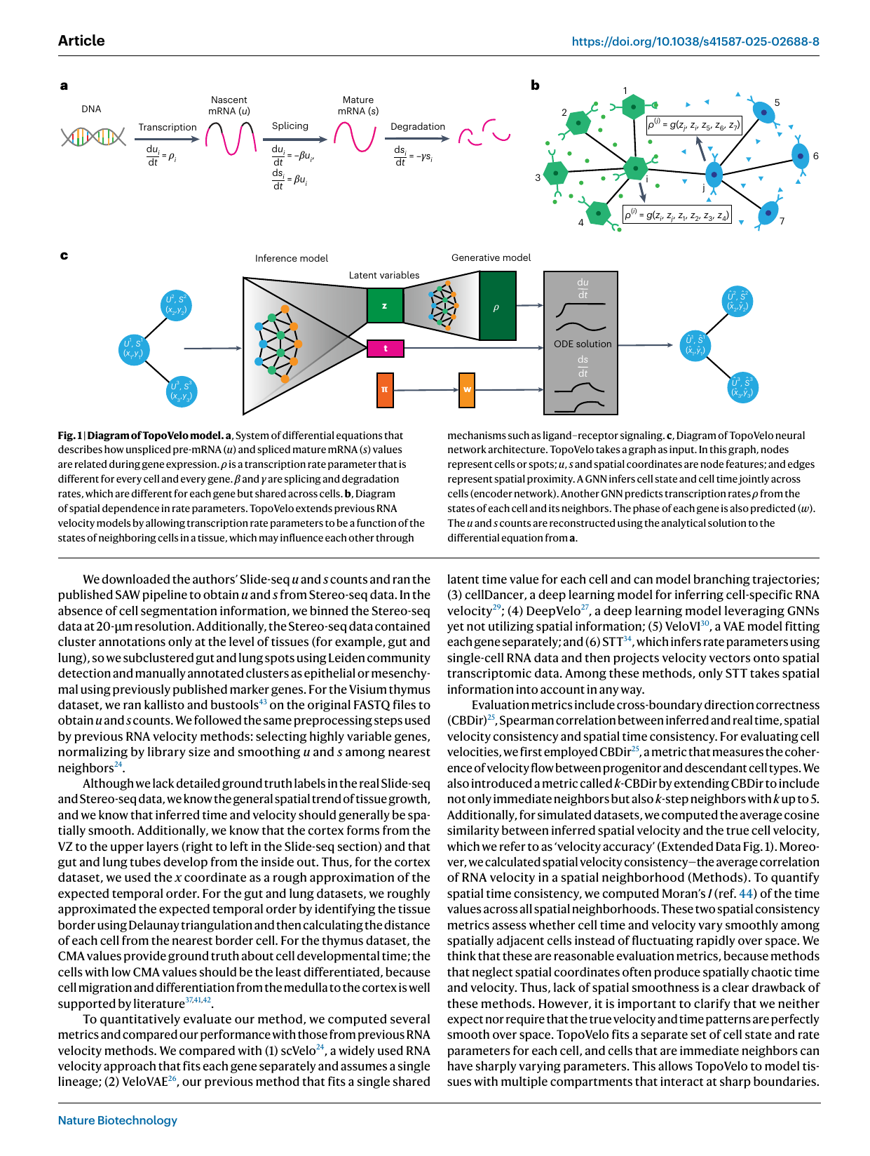
Fig 1 — TopoVelo model (Gu et al., Nat Biotechnol 2025; topovelo). The splicing ODE (a) du/dt = ρ − βu, ds/dt = βu − γs is made spatially coupled: a cell’s transcription rate ρ^(i) = g(z_i, {z_j : j∈nbr(i)}) is predicted from its own and its spatial neighbors’ states over the tissue graph (b). A graph-VAE (c) encodes cells into latent state z, cell time t, and gene phase, with a generative decoder solving the ODE — the spatial analog of RegVelo’s GRN coupling.
Two velocities
- RNA / expression velocity — change in (u, s), as in standard RNA velocity.
- Cell (migration) velocity — change in spatial position w.r.t. time, in μm/hour. This is TopoVelo’s distinctive output.
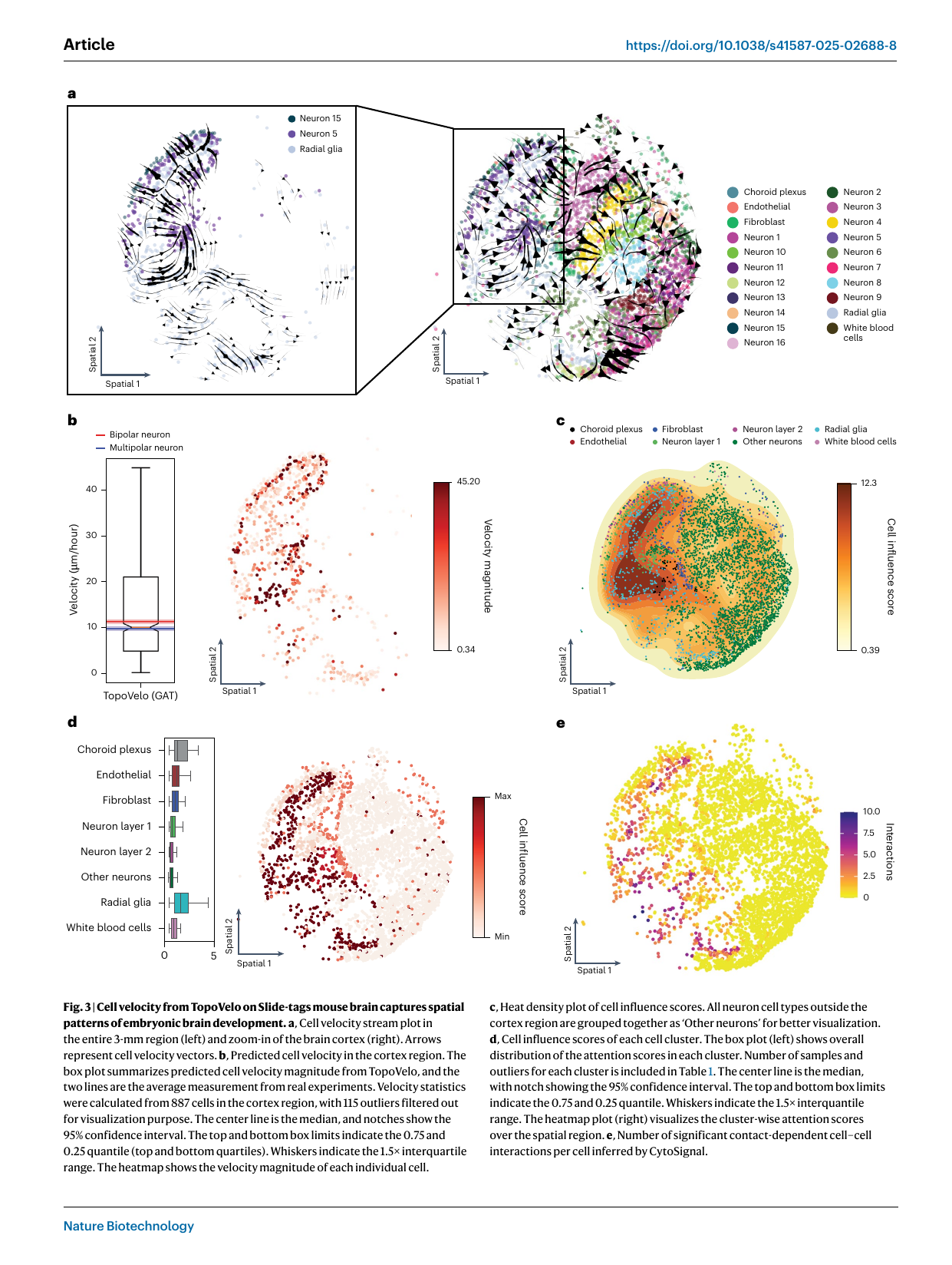
Fig 3 — cell velocity in physical units (Gu et al., Nat Biotechnol 2025; topovelo). On Slide-tags mouse brain, the cell-velocity stream plot (a) and the per-cell velocity magnitude (b) give a median ~10 μm/hour, matching the live-cell imaging bracket (9.8 ± 0.4 multipolar, 11.3 ± 0.4 μm/h bipolar neurons, overlaid lines) — the wiki’s first physical-velocity validation. The cell influence score (graph attention; c–d) is highest in radial glia and choroid plexus, co-locating with CytoSignal contact-dependent signaling (e). Note: μm is measured; the per-hour scale rests on the assumed 20 h gene cycle (see spatial-velocity, physical-time-grounding).
Physical-time scorecard
| Axis | TopoVelo |
|---|---|
| Latent time | inferred t is ordinal; converted to physical units via an assumed 20 h gene cycle |
| Rate scale | expression timescale assumed (20 h); spatial scale real (μm) → migration velocity anchored |
| External anchor | partial/indirect: μm coordinates + match to live-cell-imaging migration rates (~10 μm/h); no labeling |
| Constant rates | ρ spatially coupled; β, γ constant |
| Verdict | metric-by-assumption + external spatial validation; closest to physical units, but timescale imported not identified |
See physical-time-grounding and spatial-velocity.
Cell influence score
Graph-attention weights → a per-cell “influence score.” High-influence cells (radial glia, choroid plexus, fibroblasts; WNT-signaling cells in embryoid bodies) co-locate with ligand–receptor / contact-dependent signaling hotspots (validated via CytoSignal).
Validated on / applications
Slide-seq / Stereo-seq / 10x Visium (E15 mouse cortex, E13.5 gut & lung, human thymus), Slide-tags mouse brain, 3D Slide-seq mouse embryo (neural-tube closure via spatial- velocity divergence along A–P axis), and human embryoid bodies (Curio Seeker).
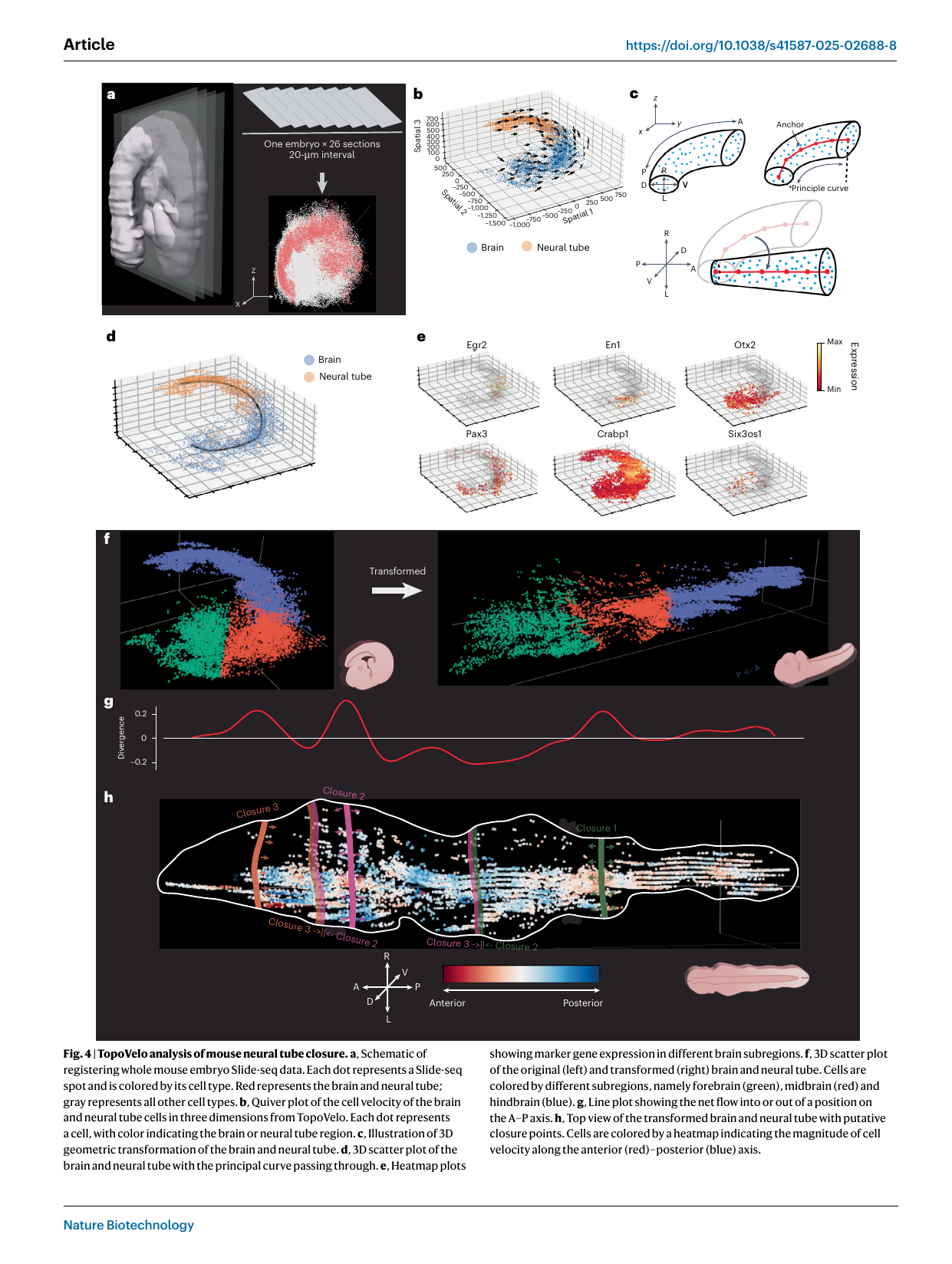
Fig 4 — mouse neural-tube closure (Gu et al., Nat Biotechnol 2025; topovelo). A whole E9.0 mouse embryo is reconstructed in 3D from 26 registered Slide-seq sections (a). TopoVelo’s 3D spatial-velocity field over brain + neural tube (b) is “unrolled” along an elastic principal curve fit to the A–P axis (c–f); the divergence of cell velocity along A–P (g) peaks at three positions matching the known closure points C1/C2/C3 (h) — annotating neural-tube closure data-drivenly from spatial velocity.
Relation to other methods
- Extends VeloVAE (adds spatial coupling).
- Spatial analog of RegVelo (grn-informed-velocity); context = neighbors vs GRN.
- Outperforms STT, Spateo, cell2fate, GASTON (prior spatial-velocity methods) and non-spatial scVelo, veloVI, DeepVelo, cellDancer on spatial consistency.
- Closest in the wiki to dynamo’s physical grounding, but via spatial units + an assumed clock rather than metabolic-labeling.
Related
spatial-velocity · VeloVAE · RegVelo · grn-informed-velocity · splicing-kinetics-ode · latent-time · physical-time-grounding · metabolic-labeling · RNA velocity · FlowVelo